Ultrasound-guided posterior tibial nerve block: why seeing the needle makes it safer
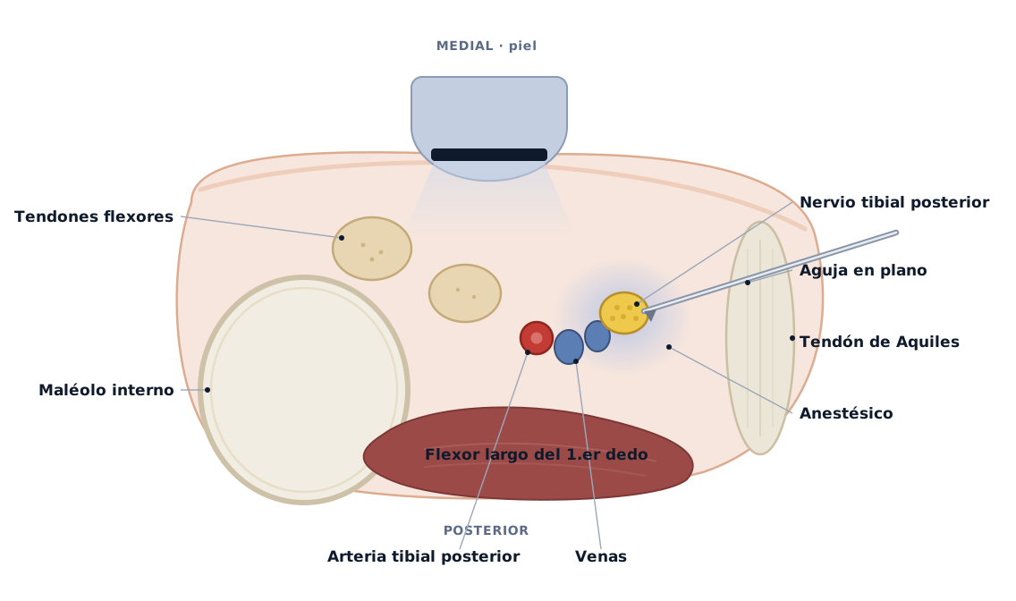
The posterior tibial nerve supplies sensation to the sole of the foot. Blocking it allows the sole and the heel to be anaesthetised for surgery or painful wound care without numbing the whole leg. It runs behind the medial malleolus, right beside the posterior tibial artery and vein. That is where the difference lies between doing it blind and doing it with ultrasound: seeing the needle, the nerve and the artery in real time lets us place the anaesthetic around the nerve without touching the vessel.
What it is and what it is for
The posterior tibial nerve block is a regional anaesthetic technique for the foot. Instead of injecting blind, guided only by skin landmarks, ultrasound locates the nerve next to the posterior tibial artery and lets us place the anaesthetic exactly where it needs to act. It is used for procedures on the sole and the heel: surgery, wound care, pain management. Its main strength is not just that it works: seeing everything on screen reduces the risks of the needle pass itself.
Why ultrasound changes the safety picture
With ultrasound, less toxicity from the anaesthetic
Registry of 20,021 patients and 25,336 nerve blocks: ultrasound guidance was associated with less local anaesthetic systemic toxicity, the most feared complication. Overall incidence was 0.87 per 1,000 blocks (95% CI 0.54 to 1.3).
Systemic toxicity occurs when the anaesthetic reaches the bloodstream, almost always through unintentionally puncturing a vessel. Seeing the needle and the artery on screen is precisely what helps avoid that. It is a rare event, so no single study "proves" it, but this registry is the best population-level signal that ultrasound adds safety.
Barrington MJ, Kluger R. Ultrasound guidance reduces the risk of local anesthetic systemic toxicity following peripheral nerve blockade. Reg Anesth Pain Med. 2013;38(4):289-99. doi:10.1097/AAP.0b013e318292669b
Safer in some respects, not in all
The ASRA evidence-based review concludes that ultrasound reduces local anaesthetic systemic toxicity and transient paralysis of the diaphragm, but does not lower the rate of neurological symptoms after a block.
It is worth saying plainly: ultrasound makes the block safer in some respects and not in others. It reduces the risk of injecting into a vessel and of toxicity, but the possibility of irritating the nerve does not disappear just because a scanner is used. The technique adds safety, it does not guarantee it, and it still calls for trained hands.
Neal JM. Ultrasound-Guided Regional Anesthesia and Patient Safety: Update of an Evidence-Based Analysis. Reg Anesth Pain Med. 2016;41(2):195-204. doi:10.1097/AAP.0000000000000295
The posterior tibial block, in the foot
It numbs the foot without weakening the leg
In day-case heel surgery, an ultrasound-guided posterior tibial nerve block 10 cm above the medial malleolus provided 6 hours of analgesia and kept foot strength intact (plantar flexion 5/5), with pain-free discharge and no complications at one month.
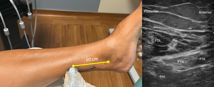
The classic alternative, blocking the sciatic nerve higher up, leaves the leg weak and increases the risk of falls. Locating the posterior tibial nerve with ultrasound lets us anaesthetise only what is needed and preserve the muscles. The image shows what appears on screen: the nerve right next to the artery, which is the structure best left untouched.
Garcia Tomas V, DeLeon AM, Johnson PA, Vargas K, MacLyman S, Chung B. Proximal Ultrasound-Guided Posterior Tibial Nerve Block for the Removal of Calcaneal Hardware. Cureus. 2023;15(6):e41047. doi:10.7759/cureus.41047
From pain of 9 to 0 in under ten minutes
In a series of patients with severe foot pain (7 to 10 out of 10), the ultrasound-guided posterior tibial nerve block brought the pain down to 0 in under 10 minutes, with no local or systemic toxicity in any case.
The technical detail is the key to safety: the needle is advanced "in plane", keeping the posterior tibial artery and vein in view, and saline is injected first to confirm that the tip sits at the nerve and away from the neighbouring structures before the anaesthetic is given. The authors sum up that, compared with relying on skin landmarks alone, ultrasound gives better pain control and fewer complications.
Burl T, Latshaw P, Dreyfuss A. Ultrasound-Guided Posterior Tibial Nerve Block for Frostbite of the Plantar Surfaces: A Case Series. Clin Pract Cases Emerg Med. 2022;6(4):272-275. doi:10.5811/cpcem.2022.7.56727
Take-home
- The posterior tibial nerve anaesthetises the sole of the foot and runs right beside the posterior tibial artery, behind the medial malleolus.
- Guiding the block with ultrasound shows the needle, the nerve and the artery at the same time: the anaesthetic goes where it should and the vessel is avoided.
- Registry evidence links ultrasound guidance with less local anaesthetic systemic toxicity, the most feared complication.
- It is not magic: ultrasound reduces some risks (injecting into a vessel, toxicity) but does not remove the possibility of irritating the nerve. It adds safety and precision, hand in hand with a well-trained technique.
At Avanza Salud we do it under ultrasound guidance
We perform ultrasound-guided anaesthetic blocks of the foot in our own clinic: we locate the nerve next to the artery and place the anaesthetic exactly where it belongs, watching the needle in real time to steer clear of what must not be touched. It is the same control we apply across the rest of our ultrasound-guided procedures. If you are due to have a procedure on your foot, or you have pain that might benefit from a nerve block, we will assess it with you and explain clearly what makes sense in your case.