Muscle tears: classification and return to sport
"Torn muscle fibres" is still the phrase most people use, but since 2013 sports medicine has worked with something more precise: the Munich classification, which separates functional injuries (no macroscopic tear) from structural ones (with a tear). The distinction matters, because it changes the prognosis, the imaging you need and how long rehabilitation takes.
Functional versus structural
The Munich consensus defined four types: type 1 (overuse-related functional disorder), type 2 (neuromuscular functional disorder), type 3 (structural injury, 3A minor partial tear / 3B moderate partial tear) and type 4 (subtotal tear or tendinous avulsion). Types 1-2 show no damage on imaging yet limit function; types 3-4 show a visible tear. A functional injury can settle in days; a 3B or type 4 needs weeks of structured rehabilitation.
The Munich classification
Four types, two clinical categories
International consensus published in Br J Sports Med: four types of muscle injury, 1-2 functional (no tear) and 3-4 structural (with a tear). Types 1-2 show no damage on imaging but do limit function; types 3-4 show a macroscopic tear on ultrasound or MRI.
The classification changes management from day one: a functional injury can settle within days on a progressive plan, whereas a 3B or type 4 tear needs weeks of structured rehabilitation and careful planning of the return to sport. Labelling the injury properly determines the imaging required, the prognosis and a realistic timeframe for competing again.
Mueller-Wohlfahrt HW, Haensel L, Mithoefer K, Ekstrand J, English B, McNally S, et al. Terminology and classification of muscle injuries in sport: the Munich consensus statement. Br J Sports Med. 2013;47(6):342-50. doi:10.1136/bjsports-2012-091448
What predicts re-injury
A recent injury multiplies the risk by almost five
Systematic review and meta-analysis of 78 studies, 8,319 injuries and 71,324 athletes: older age (SMD 1.6; p=0.002); any previous hamstring strain injury RR 2.7 (p<0.001); a recent hamstring strain injury RR 4.8 (p<0.001), the strongest risk factor of all; previous ACL injury RR 1.7; previous calf injury RR 1.5.
The clinical message: the main risk factor for the next injury is the last one, particularly when rehabilitation was left unfinished. And one finding worth holding on to before over-reading a scan: the authors conclude that recurrence risk is judged better from clinical data than from the MRI of the index injury.
Green B, Bourne MN, van Dyk N, Pizzari T. Recalibrating the risk of hamstring strain injury (HSI): A 2020 systematic review and meta-analysis of risk factors for index and recurrent hamstring strain injury in sport. Br J Sports Med. 2020;54(18):1081-1088. doi:10.1136/bjsports-2019-100983
The prevention with the strongest evidence: the Nordic hamstring
Programmes including the Nordic exercise cut hamstring injuries by up to 51%
Systematic review and meta-analysis of five studies in footballers: programmes including the Nordic hamstring exercise achieved a pooled IRR of 0.490 (95% CI 0.291-0.827; p=0.008). Teams that ran the programme reduced hamstring injuries by up to 51% in the long run compared with teams without a prevention programme.
The limitation the authors acknowledge is adherence. Later studies in professional leagues report low compliance, which dilutes the effect in the real world. It works if it is done, not if it is merely prescribed. Rehabilitation after a muscle injury still runs in phases (pain control → strength → functional loading → sport-specific movement → competition), and the return to sport is decided on functional criteria, not by the calendar and not by how the scan looks.
Al Attar WSA, Soomro N, Sinclair PJ, Pappas E, Sanders RH. Effect of Injury Prevention Programs that Include the Nordic Hamstring Exercise on Hamstring Injury Rates in Soccer Players: A Systematic Review and Meta-Analysis. Sports Med. 2017;47(5):907-916. doi:10.1007/s40279-016-0638-2
How many days each type actually takes
Six days against sixteen, and thirty-two with a moderate tear
Validation of the Munich classification across 393 thigh injuries in 31 elite European clubs. Functional injuries cost a median of 6 days; structural injuries, 16. Within the structural group: 3A, 13 days; 3B, 32 days; type 4, 60 days.
This is the finding that makes the classification useful to an athlete, because it puts numbers on the question everyone asks on day one. They should be read as medians for a group rather than as a personal date: the interquartile range for 3B injuries is 24 days, so two injuries carrying the same label can end up weeks apart.
Within the functional injuries there were no differences between subtypes, all falling between four and eight days. Only 5% of them progressed to a structural injury within two months, which supports not over-resting a complaint without a tear.
Two limits the authors themselves acknowledge. The type 4 group contained only five injuries, so its 60 days is indicative at best. And the medical team that classified each injury was the same one that later cleared the player, with no independent assessor, which can reinforce the agreement between label and timeframe.
Ekstrand J, Askling C, Magnusson H, Mithoefer K. Return to play after thigh muscle injury in elite football players: implementation and validation of the Munich muscle injury classification. Br J Sports Med. 2013;47(12):769-74. doi:10.1136/bjsports-2012-092092
What to check before clearing an athlete
Tenderness on palpation at clearance quadruples re-injury
Twelve-month follow-up of 64 athletes cleared after a hamstring injury, with 17 re-injuries. Remaining tenderness on palpation multiplied the risk by 3.95 (95% CI 1.38-11.37). Deficits in active knee extension and in isometric flexion strength also counted, as did the number of previous injuries.
What sets this study apart is that it measured the variables at the moment of clearance and then waited to see who broke down, rather than describing which criteria are used out of habit. That makes it one of the few references connecting a specific examination to what happens next.
No variable from the baseline MRI was associated with re-injury, a result worth remembering before resting a clearance decision on imaging.
It needs weighing carefully: with 17 re-injuries and four variables in the model, the confidence intervals are wide and overfitting is a real possibility. The strength and range-of-motion deficits were also associated with small increases per unit of deficit, so they only carry clinical weight when the difference between legs is appreciable, not for a minimal asymmetry.
De Vos RJ, Reurink G, Goudswaard GJ, Moen MH, Weir A, Tol JL. Clinical findings just after return to play predict hamstring re-injury, but baseline MRI findings do not. Br J Sports Med. 2014;48(18):1377-84. doi:10.1136/bjsports-2014-093737
No clearance criterion is validated, but some hold up better than others
Systematic review of 135 studies on return-to-sport criteria in footballers. The most widely used for hamstrings are strength symmetry between legs and absence of pain during sport-specific movements. The ones reaching the highest certainty of evidence are active range of motion and the athlete's own readiness.
The uncomfortable message from this literature is that no validated battery guarantees a safe return. Earlier reviews had already pointed out that the usual criteria, no pain, comparable strength and flexibility, and medical clearance, are applied almost universally without ever having been tested against re-injury.
That the athlete's own readiness appears among the better-supported criteria is not a footnote. Going back while feeling unsure is clinical information, not weakness, and it belongs in the decision alongside strength and mobility.
The review maps what is used and how well it is supported methodologically; it does not show that applying those criteria reduces re-injury. It includes male footballers only, so transferring it to other populations is a reasonable but untested extrapolation.
Pecci J, van Dyk N, Myer GD, Sañudo B. Return to Sport Criteria After Muscle Injury in Male Football Players: A Systematic Review with Evidence Certainty Synthesis. Sports Med. 2026;56(6):1433-65. doi:10.1007/s40279-026-02404-9
What imaging adds, and what it does not
MRI added 2.8% to what the examination already said
In 180 athletes assessed within the first five days, a model based on history and physical examination alone explained 29% of the variance in time to return to sport. Adding MRI took it to 31.8%. The authors conclude there is no justification for requesting it routinely in acute hamstring injury.
It is worth reading the other half of the result too: even with examination and MRI combined, two thirds of the variance remains unexplained. Giving an exact date on day one is imprudent with any test in hand.
Other work points the same way. MRI classification systems explain between 8% and 12% of the timeframe, and the size of the oedema between 6% and 12%. In a series of 53 athletes, 89% still showed abnormalities on imaging on the day they were cleared, and they competed anyway: waiting for the MRI to normalise is not a requirement.
Imaging does have another role. In the largest cohort to date, with 330 injuries, involvement of the myotendinous junction on the initial MRI was associated with a threefold risk of re-injury within a year. In other words, imaging says more about the risk of breaking down again than about the date of return, and those are two different questions.
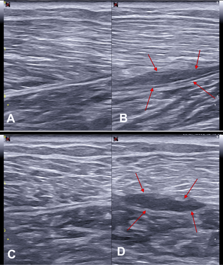
As for ultrasound, the available evidence places it on a par with MRI in the acute phase, with 85% sensitivity and 84% specificity using MRI as the reference, plus the advantage of a dynamic examination in the same consultation. Its weak point is injuries involving the tendon, where agreement between the two tests falls to 60%, and long-term follow-up.
Wangensteen A, Almusa E, Boukarroum S, Farooq A, Hamilton B, Whiteley R, et al. MRI does not add value over and above patient history and clinical examination in predicting time to return to sport after acute hamstring injuries. Br J Sports Med. 2015;49(24):1579-87. doi:10.1136/bjsports-2015-094892
Take-home
- The Munich classification separates functional injuries (1-2) from structural ones (3-4) and guides the prognosis.
- A recent injury is the strongest risk factor for the next one (RR 4.8).
- The Nordic hamstring exercise cuts hamstring injuries in footballers by up to 51%, provided it is actually done.
- A functional injury costs around 6 days and a structural one 16; with a moderate partial tear (3B) the median rises to 32 days.
- Still being tender on palpation on the day of clearance quadruples the risk of re-injury.
- MRI adds little to the examination for estimating the timeframe, though it does say something about the risk of breaking down again.
- Return to sport should rest on clinical and functional criteria, not on the calendar and not on imaging alone.
At Avanza Salud we can help
We manage muscle injuries with a comprehensive approach: clinical and ultrasound assessment to classify the injury properly, a phased return-to-sport programme (pain control → strength → functional loading → sport-specific movement), specific preventive exercise including the Nordic hamstring, and a return to sport based on measurable criteria. If you have been injured, or have broken down more than once in the same muscle group, it is worth reviewing the whole plan before competing again.